Workplace and personal injuries often affect joins throughout the body, especially ones in more mobile areas like the spine, legs, arms, and shoulders.
If there is a need to visualize, diagnose, or treat damaged joints to determine the extent of an injury, an arthroscopy may be performed. It’s a procedure performed in a way that’s less invasive and often safer for the patient.
Diagnostic Arthroscopy
When joints are injured after a slip-and-fall accident or while on-the-job, the first attempt at making a diagnosis usually involves X-rays, CT scans, or MRI scans. But if image tests alone aren’t providing enough information about what may be going on internally, an arthroscopy may be recommended. Common joints examined arthroscopically include knees, shoulders, elbows, ankles, hips, and wrists. The goal with a diagnostic arthroscopy is to determine the full extent of the injury by examining very detailed, real-time internal images.


Arthroscopic Surgery
Surgery can usually be performed with minimally invasive arthroscopic techniques when the damage to the joint is fairly localized. This being said, there are times when traditional open surgery is more appropriate. However, some surgical procedures done arthroscopically have become routine. One example of this is the meniscal tears in the knee, which are often successfully treated with arthroscopic surgery. Specific reasons for arthroscopic surgery may also include:
- Inflammation in the lining of joints (synovitis)
- Rotator cuff tendon tears
- Carpal tunnel syndrome
- Issues with loose bodies of cartilage and/or bone
Preparations for the Procedure
Prior to performing an arthroscopy, certain medications that increase the risk of bleeding may need to be temporarily stopped. Depending on what type of anesthesia will be used, patients may also be instructed not to eat solid foods for about 8 hours before the procedure is scheduled to be performed.
How an Arthroscopy Is Performed
During the procedure, a patient is positioned in way that allows convenient access to the affected joint. If it’s appropriate to do so, the affected limb may be placed in a special positioning device to prevent movement. A tourniquet is sometimes used to reduce issues with blood loss. Also, the area around the joint may be injected with a sterile fluid to expand the area surrounding the affected joint.
The procedure involves a small incision for the insertion of the lighted arthroscope, which also has a lens attached to it so images can be viewed on a monitor. If surgery is performed, additional small incisions (accessory incisions) will be made for specialized designed instruments to be inserted to perform the intended procedure. When an arthroscopy is done, the incisions are covered with bandages or dressing. In some cases, “liquid stitches” or similar incision closure methods may be used.
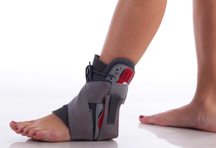
An arthroscopy is generally considered to be a safe procedure for both diagnostic and treatment purposes. Aftercare will depend on the reason for performing an arthroscopy in the first place. Follow-up care might include physical therapy or therapeutic exercises, the temporary use of medication to relieve pain or reduce inflammation in the affected area, the RICE method to allow the affected joint to heal, or the temporary use of crutches, splints, or slings. If the procedure was done for diagnostic purposes, other types of surgery may be performed.

